 |
Science Frontiers ONLINE No. 37: Jan-Feb 1985 |
|
|
Parasites may reprogram host's cell
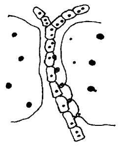
The long, segmented filament shown in the illustration consists of the cells of a parasite that preys on the cells of red algae. Two such cells abut the parasitic filament. The small black circles are parasitic cell nuclei which, when confronted with a red alga cell, become wrapped in small "conjuctor cells" which are then somehow transferred to the host cell on the right. The actual transfer involves the formation on the host cell wall of a sort of dimple called a "secondary pit connection." (Why this forms is not known.) Once inside the host cell, the parasite nucleus and/or the cytoplasm transferred with it dramatically reprograms host cell operations. The host cell shifts into highgear food production, enlarging up to twenty times its normal size. The host cell wall thickens, its nuclei (large black circles) and chloroplasts multiply. Adjacent cells (left) remain unaffected.
(Lewin, Roger; "New Regulatory Mechanism of Parasitism," Science, 226:427, 1984.)
Comment. It is one thing for external stresses to stimulate cell reprogramming, but quite another when another species inserts its programs into those of the host. Is this another way of producing "hopeful monsters?"
 | A parasitic filament growing between host cells, showing how parasite nuclei are first enclosed in bud-like conjunctor cells and then inserted into the host cell. |